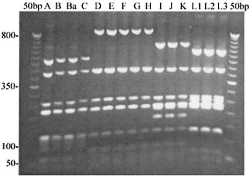
FIG. 3.
PmpE digested with HinfI restriction endonuclease shows segregation of ocular, urogenital, and LGV serovars. An 8-μl aliquot of the pmpE PCR products was digested with HinfI endonuclease, and restriction fragments were separated on a 2% agarose gel. Serovars are indicated above their respective lanes. Numbers on the left are size standards in base pairs.

