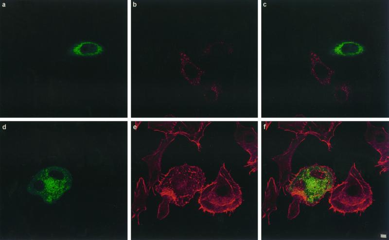
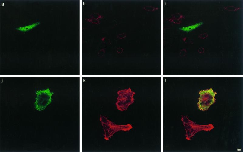
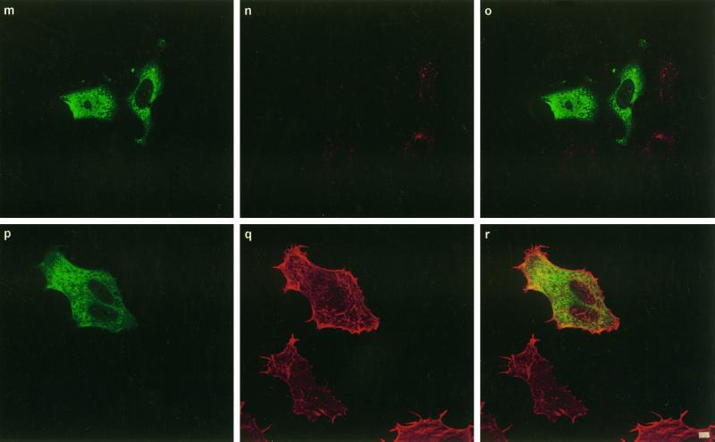
Figure 3.
Confocal immunofluorescence microscopic analysis of coated vesicle inhibition in HEp-2 cells by transfection of DNAs encoding ED95/295, dyn-K44A, and int-SH3: effects on transferrin internalization and CNF1 activity. Transfected cells are identified either by GFP tags, ED95/295 (a and d) and int-SH3 domain (m and p), or by the HA tag using mAb 12CA5 anti-HA followed by binding of an anti-mouse FITC-labeled antibody (g and j). Cells were controlled for transferrin endocytosis by incubation with 2 mg/ml Texas Red-labeled transferrin for 90 min at 37°C (b, ED95/295; h, dyn-K44A; n, int-SH3 domain), (c, i, and o) Merged images of a and b, g and h, and m and n, respectively. Cells transfected with ED95/295 (e), dyn-K44A (k), or int-SH3 (q) were incubated with 10−9 M CNF1 for 90 min. Bafilomycin A1 was then added to block further entry of the toxin (see MATERIALS AND METHODS), and cells were incubated for an additional period of 24 h with bafilomycin A1 to develop the CNF1 phenotype consisting of cell spreading, membrane ruffling, stress fiber formation, and multinucleation (clearly shown in e, k, and q). Cells were then stained for F-actin with Texas Red-phalloidin (e, k, and q). (i, f, and r) Merged images of d and e, j and k, and p and q, respectively. Bars, 2 mm.


