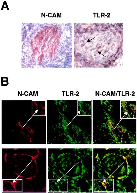
FIG. 3.
TLR2 is expressed in vivo on Schwann cells in leprosy lesions. (A) Representative sections of skin biopsy specimens from leprosy patients showing the expression of NCAM and TLR2. Arrows indicate cells with wavy nuclei, characteristic of nerve cells. (B) Two-color immunofluorescence staining of skin lesions from leprosy patients. Cryostat sections of skin biopsy specimens were stained with anti-NCAM (red, left panels) and anti-TLR2 (green, center panels) antibodies. The merge of the two images (right panels) showed colocalization of NCAM and TLR2. The presence of nerves in small biopsy specimens was variable. We analyzed almost 20 patients representing the spectrum of leprosy; the lesions shown were from a patient with erythema nodosum leprosum, a reaction in lepromatous patients. The insets duplicate and highlight the doubly positive cells typical of Schwann cells (arrows). Original magnification, ×630.

