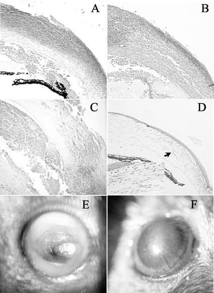
FIG. 1.
Histological and clinical examination of mouse corneas infected with P. aeruginosa. Histological sections are stained with hematoxylin and eosin. The magnification of all sections is ×200, except for the inset in panel D, which is at ×400. (A) Wild-type mouse at 24 h postchallenge; (B) IL-10−/− mouse at 24 h postchallenge; (C) wild-type mouse at 7 days postchallenge; (D) IL-10−/− mouse at 7 days postchallenge (the arrow indicates the area shown at higher magnification in the inset); (E) wild-type mouse at 7 days postchallenge; (F) IL-10−/− mouse at 7 days postchallenge showing increased vascularization.

