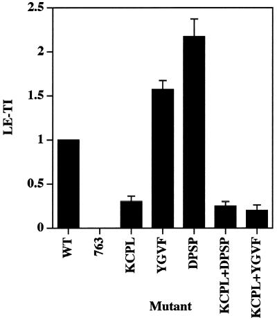
Figure 3.
Quantitation of targeting of HRP-P-selectin chimeras to late endosomes. PC12 cells expressing the chimeras indicated were homogenized and fractionated on 1–16% Ficoll velocity gradients followed by recentrifugation on 0.9–1.85 M sucrose equilibrium gradients for separation of late endosomes. LE-TIs were then calculated by normalizing the amount of HRP activity within the endosomal peak both for organelle recovery as judged by NAGA activity and for the expression level. Each bar represents the mean ± SE of three independent experiments.

