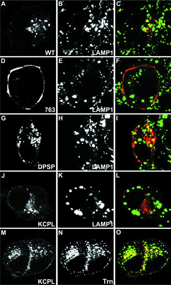
Figure 5.
Immunofluorescent localization of internalized HRP-P-selectin chimeras and endosomal markers in PC12 cells. Cells transiently expressing wild-type ssHRPP-selectin (A–C), tailless ssHRPP-selectin763 (D–F), ssHRPP-selectinDPSP (G–I), or ssHRPP-selectinKCPL (J–O) grown on poly-l-lysine-coated coverslips were washed with serum-free medium containing 1% BSA and fed with 50 μg/ml Trn (J–O) and/or with 5 μg/ml 2H11 (A–O) for 2 h at 37°C. Cells were then fixed, permeabilized, and stained as described in MATERIALS AND METHODS. 2H11 (A, D, G, J, and M) was visualized with Texas Red-conjugated goat anti-mouse secondary antibody; LAMP1 (B, E, H, and K) was immunodetected with rabbit polyclonal anti-LAMP1 followed by FITC-conjugated goat anti-rabbit secondary antibody; and Trn (N) was detected with rabbit polyclonal anti-Trn followed by FITC-conjugated goat anti-rabbit secondary antibody. Color images show the merger of both channels.

