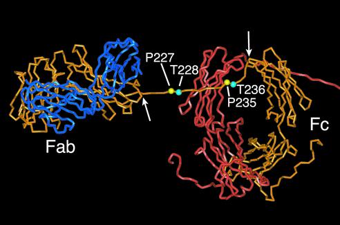
FIG. 8.
Partial view of a molecular model of human IgA1 highlighting the hinge residues investigated in this study. Coordinates were taken from Brookhaven Protein Data Bank as PDB code 1iga (6). For clarity, one entire heavy chain (orange), one entire light chain (blue), and the Fc and partial hinge of the second heavy chain (red) are shown (the remainder of the heavy chain and the second light chain lie out of field to the right of the picture). Proline residues Pro227 and Pro235 are highlighted in yellow, and Thr228 and Thr236 are shown in cyan. This model, based on X-ray and neutron scattering data (6), predicts that the hinge of each heavy chain crosses over the CH2 domain of the other heavy chain and adopts an extended conformation. The arrows indicate residues Val222 and Cys241, the points of constraint at each end of the hinge region, between which the hinge is assumed to be relatively mobile.

