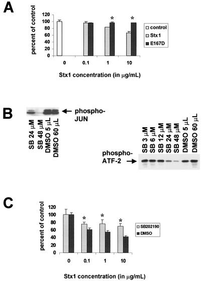
FIG. 5.
Effect of SB202190 on Stx1-induced cell death and Stx1-induced p38 and JNK activation. (A) After treatment of HCT-8 cells for 72 h with various concentrations of Stx1 or Stx1E167D, cell viability was measured by MTT assay expressed as percentage of control cell viability. (B) Effects of various doses of SB202190 on Stx1-induced p38 and JNK activation in HCT-8 cells. Briefly, cells were preincubated with doses of SB202190 ranging from 3 to 48 μM, or equal amounts of the SB202190 diluent DMSO, and then exposed to Stx1 at 1 μg/ml. Cell extracts were obtained and assessments of p38 and JNK activation were performed as outlined in Materials and Methods. (C) After treatment of HCT-8 cells for 48 h with various concentrations of Stx1 with and without the p38 inhibitor SB202190, cell viability was measured by MTT assay and expressed as percentage of control cell viability. In panels A and C, optical density measurements for control cells were averaged, and then the optical density (at 540 nm) reading for each well in the microtiter plate was expressed as a percentage of the control average, including the individual control measurements. The percentages were then averaged for each group, and data are expressed as a mean ± standard deviation. For each condition, n = 3 wells. In panels A and C, * denotes a P value of <0.05 compared with control average.

