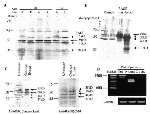
Figure 2.

Detection of chondrocyte expression of receptor for advanced glycation end products (RAGE) by immunoblotting and reverse transcription–polymerase chain reaction (RT-PCR). A, Lysates were prepared from cells (obtained from 2 human donors) freshly isolated (culture −) from human knee (K) or ankle (A) articular cartilage or from cells isolated from the same joints but cultured in monolayer for 5–7 days (culture +). Equal amounts of total protein (10 μg) were run in each lane and immunoblotted with an anti-RAGE monoclonal antibody (MAB5328). Arrows to the right of the blot indicate potential RAGE isoforms. It is not known if the band just below 75 kd is related to RAGE. Molecular weight markers are shown at the left. B, Cell lysate samples were prepared from cells cultured in monolayer for 5 days (control) and from cells from the same donor that were transfected with a construct that expresses RAGE. One-half of the lysate sample was treated with glycopeptidase F prior to sodium dodecyl sulfate–polyacrylamide gel electrophoresis and immunoblotted for RAGE with the monoclonal antibody. C, Immunoblotting was performed with the monoclonal antibody using a cell lysate from monolayer-cultured cells and from a direct cartilage extract. The same blot was stripped and reprobed with a polyclonal antibody (C-20) to the RAGE C-terminus. D, RT-PCR was performed using RNA isolated from primary monolayer cultures, which was amplified using 3 different RAGE primer sets and GAPDH primers as a control. The RAGE primers were designed to amplify regions coding for full-length (full), N-truncated (N-trunc), and C-truncated (C-trunc) RAGE.
