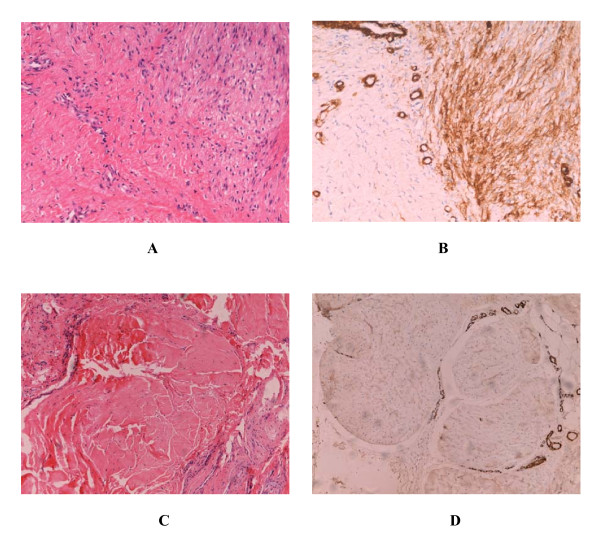
Figure 2.
Histopathological and immunohistochemical features of palmar fibromatosis. Three distinct histological phases can be observed in the patient 9. The right part of Panel A (Hematoxylin and eosin × 200) shows proliferative phase of the lesion and the left part shows evolutional phase. Panel B (ABC × 200) shows the expression of α-SMA. Spindled cells of proliferative stage formed nodule and strongly expressed α-SMA. In evolutional stage, a majority of myofibroblasts were replaced by fibroblasts, and spindled cells were separated by the collagen. Panel C (Hematoxylin ane eosin × 200) shows residual phase of the lesion, and panel D (ABC × 200) shows the expression of α-SMA. Spindled cells disappear and are substituted by amounts of dense collagen. Except smooth muscle cells of blood vessels, α-SMA is negative.

