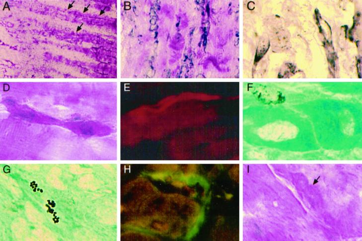
Figure 7.
A single treatment with l-NAME 30 min before injury affects dystrophic muscle regeneration. (A) Low magnification (H&E) shows a large crush region (just to the left), an adjacent region of new myotubes (arrows), and surviving fiber segments (to the right). (B) Many large new myotubes adjacent to the crush. (C) Elongated mononuclear cells and myotubes are m-cadherin+. (D) An elongated crimson cell is binucleate and located in the satellite position on a surviving fiber segment. (E) A new myotube extends from a surviving segment and contains devMHC (Texas Red fluorescence). (F) A BrdU+ nucleus next to new myotubes with unstained central nuclei. (G) A new myotube contains apoptotic BrdU+ nuclear fragments. (H) Large c-met+ satellite cells (FITC) on HGF/SF+ fibers (Texas Red) in LTA. (I) A large satellite cell (arrow) with granular cytoplasm (H&E) on an LEDL fiber has less prominent margins than in undamaged normal muscles (see Figure 5, J and K). Original magnification in A, ×13; in B, ×130; in C–I, ×330.

