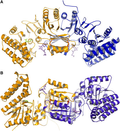
Figure 1.
Structure of AK1.
(A) Ribbon diagram of the dimer of AK1 in complex with Lys and SAM. One monomer is colored in orange and the other in blue. SAM molecules are shown in violet. Lys molecules found in the active site and in the regulatory binding site are indicated in green and red, respectively. This diagram, as well as the following diagrams of structures, was produced with the program PyMol (http://pymol.sourceforge.net/).
(B) A 90° rotated view, with respect to the horizontal axis, of the dimer showing that the regulatory domains make a core of 16 strands containing two perpendicular sheets of eight strands surrounded on both sides by four helices exposed to the solvent and flanked by the two catalytic domains.

