Figure 3.
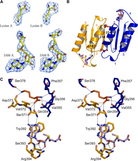
Lys and SAM Binding Sites of ACT1.
(A) Electron density maps around the Lys and SAM molecules bound to ACT1 of monomers A and B.
(B) Lys and SAM binding sites. Two equivalent Lys binding sites are located at the interface of two ACT1 domains. ACT1 of monomer A (ACT1A) and ACT1 of monomer B (ACT1B) are colored in orange and blue, respectively. SAM binding sites are located between loops 2 and 4 of each ACT1.
(C) Stereo view of the interactions of Lys and SAM molecules with amino acid residues belonging to ACT1A (residues depicted in orange sticks) and ACT1B (residues depicted in blue sticks). Bond lengths <3.4 Å are indicated. Lys and SAM molecules are depicted as red and violet sticks, respectively.

