Figure 1.
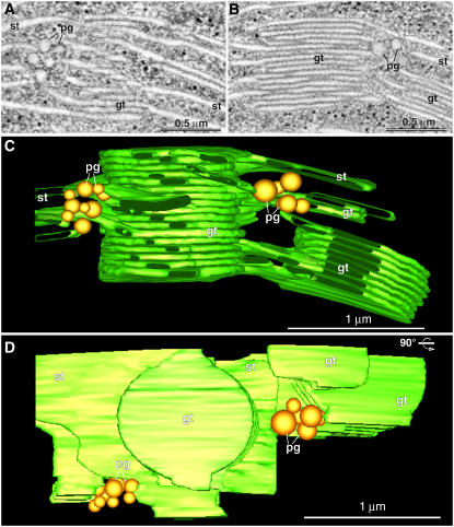
Plastoglobule Location in Relation to the Thylakoid Membranes.
(A) and (B) Two composite tomographic slice images (five superimposed serial 2.2-nm slices) of grana thylakoids (gt), stroma thylakoids (st), and plastoglobules (pg) in an intact, isolated spinach chloroplast.
(C) and (D) Tomographic model of the grana stack shown in (A) and (B) as seen in a side view (C) and a top-down view (D) ([C] rotated 90°). Note that in (D), the top-most stoma thylakoid has been removed to provide a clearer view of the clusters of plastoglobules that are associated with areas of high curvature of the stroma and grana thylakoids.

