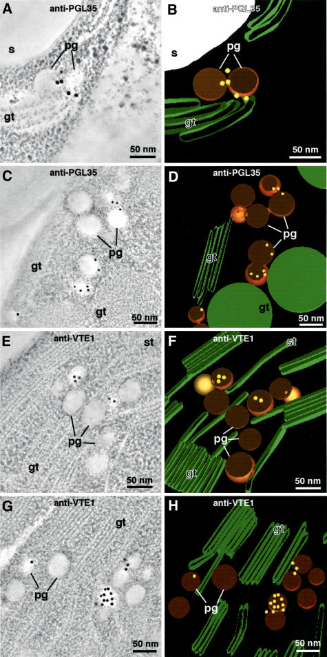
Figure 6.
PGL35 and VTE1 Immunogold Labeling.
Tomographic slice image views of plastoglobules in Arabidopsis chloroplasts immunolabeled with anti-PGL35 ([A] and [C]) and anti-VTE1 ([E] and [G]) antibodies, and corresponding tomographic models depicting the immunogold labels in a three-dimensional context ([B] and [D] and [F] and [H], respectively). The tomographic images are of 20 superimposed 2.2-nm-thick slices. Note that whereas most of the gold label appears to be associated with the coat layer of the cross-sectioned plastoglobules ([A] to [H]), one of the labeled plastoglobules in (G) exhibits labeling across its surface. As demonstrated in Figure 7, this surface corresponds to the inner surface of the plastoglobule coat monolayer. gt, grana thylakoid; pg, plastoglobule; s, starch; st, stroma thylakoid.

