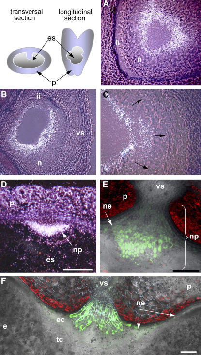
Figure 3.
Localization of jekyll Expression in Developing Caryopses.
(A) and (B) jekyll expression patterns in the nucellar tissues shortly after pollination (A) and 2 DAF (B). Hybridization sites are visualized as a white signal.
(C) Expansion of expression toward the integuments (arrows).
(D) The nucellar projection at 4 DAF.
(E) and (F) A gradient of jekyll expression within the nucellar projection 6 and 8 DAF; pjekyll:GFP (see Methods).
e, endosperm; ec, endospermal cavity; es, embryo sac; ii, integuments; n, several layers of nucellar cells; ne, nucellar epidermis; np, nucellar projection; p, pericarp; tc, transfer cells, vs, vascular tissues. Bars = 120 μm.

