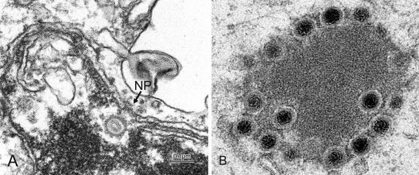
FIG. 1.
(A) Capsid of Epstein-Barr virus in front of the nuclear pore (NP) within the nucleus, showing folding of the nuclear membrane. Courtesy of Dr. V. Kushnaryov, Department of Microbiology and Molecular Genetics, Medical College of Wisconsin, Milwaukee. (B) Cytoplasmic capsid-tegument accumulation in gE-deleted bovine herpesvirus 1-infected cells.

