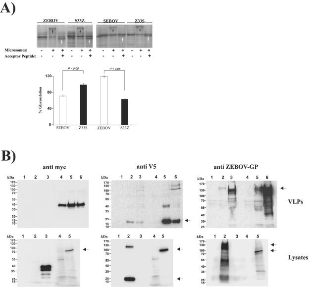
FIG. 8.
The signal peptide determines EBOV-GP interactions with the secretory pathway and is cleaved off during GP biogenesis. (A) The open reading frames encoding ZEBOV-GP, SEBOV-GP, Z33S-GP, and S33Z-GP were transcribed and translated in the presence and absence of ER-derived microsomes and an acceptor peptide, an inhibitor of glycosylation. The results of SDS-PAGE analysis of the reaction products from a representative experiment are shown in the top panel. The amounts of glycosylated reaction products were quantified by densitometry (bottom panel). The percent glycosylation represents the band intensity of the glycosylated fraction normalized to that of the precursor. The results of three independent experiments are shown. Error bars indicate SEM. (B) Western blot analysis of GP expression in cellular supernatants and purified fVLPs (top panels) or in in vitro transcription/translation reactions (bottom panels). The expression of ZEBOV- and SEBOV-GP variants harboring an N-terminal Myc and a C-terminal V5 antigenic tag was assessed by employing the indicated detection reagents. (Top panels) Supernatants of cells transfected with pcDNA3.1 (lane 1), SEBOV-GP (lane 2), or ZEBOV-GP (lane 3) or purified fVLPs produced upon cotransfection of Myc-tagged VP40 with pcDNA3.1 (lane 4), SEBOV-GP (lane 5), or ZEBOV-GP (lane 6). (Bottom panels) Cells transfected with pcDNA3.1 (lane 1), ZEBOV-GP (lane 2), Myc-tagged VP40 (lane 3), in vitro transcribed and translated pcDNA3.1 (lane 4), or in vitro transcribed and translated ZEBOV-GP (lane 5). Black arrows indicate bands corresponding to GP.

