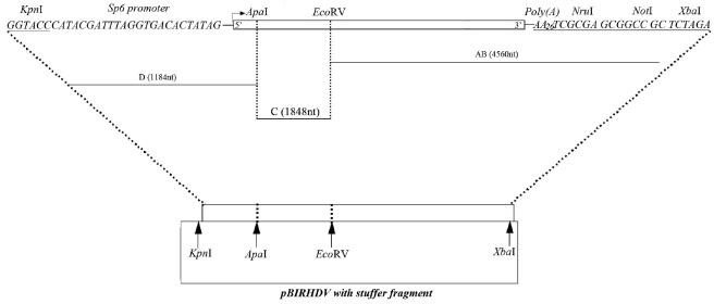
FIG. 1.
Schematic diagram of steps used in the construction of a full-length cDNA clone of RHDV. At the 5′ end of the genome, the KpnI restriction site and an Sp6 RNA promoter were fused to the genome. The arrow indicates the transcription start site of Sp6 RNA polymerase. Downstream of the 3′ untranslated region, a poly(A) tail of 27 A's and the restriction sites NruI, NotI, and XbaI were inserted. The complete viral genome was divided into three fragments flanked by unique restriction sites, represented by the horizontal lines labeled AB, C, and D. The length of each fragment is indicated in parentheses. As shown at the bottom of the figure, fragments AB, C, and D were cloned into the pBluescript II SK+ vector in the order AB to D.

