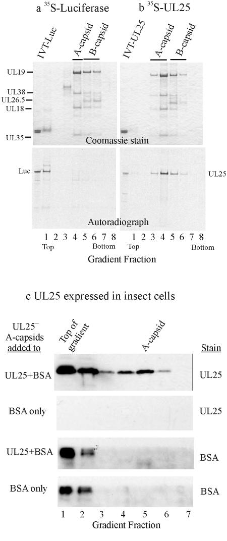
FIG. 5.
Analysis of UL25-negative capsids after exposure to UL25 in vitro. Pooled KUL25NS A and B capsids were allowed to bind [35S]Met-labeled UL25 and then purified by sucrose gradient centrifugation. Gradient fractions were then analyzed by SDS-PAGE followed by Coomassie staining (a and b, top panels) and autoradiography (bottom panels). Note that both A and B capsids bound UL25 (b) whereas neither bound the control protein luciferase (Luc) (a). IVT, in vitro transcription-translation. (c) Results obtained when KUL25NS A capsids were allowed to bind UL25 present in insect cell extracts. After incubation to permit binding to occur, capsids were purified by sucrose gradient centrifugation. Gradient fractions were then analyzed by SDS-PAGE followed by immunoblotting for UL25 (top panels) or BSA (bottom panels). All three analytical procedures were performed with the same blot. Note that capsids bound UL25 but not the control protein BSA.

