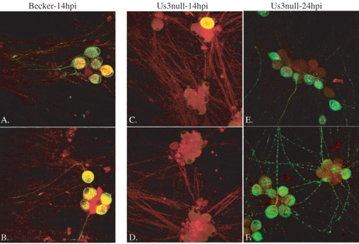
FIG. 8.
Analysis of axon-mediated infection of cell bodies. Infection was initiated in the N compartment and followed for 14 or 24 h. At these times, cells were fixed with 2% paraformaldehyde and stained with the lipid marker DiI (red) to mark all neuronal membranes and with antibodies specific for gE (green). PRV Becker (A and B)- and PRV 813 (Us3 null) (C and D)-infected cells are shown at 14 h postinfection (pi). PRV 813 (Us3 null)-infected cells are shown at 24 h pi (E and F).

