FIG. 5.
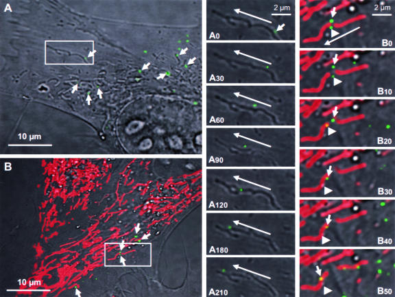
Live-cell tracking of VSV fluorescent nucleocapsid movement in infected cells. (A) BHK-21 cells were infected with VSV-PeGFP at an MOI of 10, and at 2 hpi, the culture dish was transferred to a 37°C chamber with 5% CO2 and observed under an inverted laser scanning microscope. A single infected cell and a small area (rectangular box) containing one fluorescent nucleocapsid were observed with time. Arrows in this panel identify some nucleocapsids that are in close association with mitochondrion-like structures. Panels A0 to A210 are close-up images of the small area showing the movement of a nucleocapsid (small arrow in A0) with time from the beginning (A0) to 30 s (A30), 60 s (A60), 90 s (A90), 120 s (A120), 180 s (A180), and 210 s (A210) of image recording. The direction of movement of the nucleocapsid (long arrows) toward the cell periphery is shown. (B) Live-cell tracking of nucleocapsids in infected cells stained with MitoTracker Red, which specifically stains mitochondria. The experiment was performed as described for panel A except that the infected cells were treated with MitoTracker Red for 30 min prior to image recording. Arrows identify some nucleocapsids that are in close association with red-stained mitochondria. Panels B0 (beginning) to B50 (50 s) are close-up images of the area boxed in panel B and represent the images recorded at times in seconds, as described for panels A0 to A210. Two fluorescent nucleocapsids (identified by small arrow and arrowhead) are seen moving in close association with mitochondria (red). The long arrow shows the direction of movement of the nucleocapsids toward the cell periphery.

