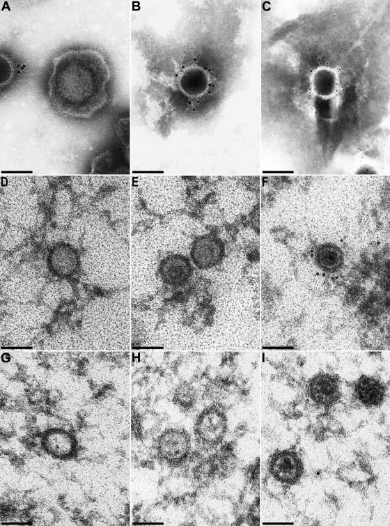
FIG. 8.
Immunolabeling of purified virions. Purified virion preparations and capsids in ultrathin sections were analyzed by immunoelectron microscopy after incubation with UL25 antiserum and gold-conjugated secondary antibodies. (A to C) Results from analysis of negatively stained purified virion preparations. (D to F) Intranuclear A, B, and C capsids in PrV-Ka-infected RK13 cells. (G to I) Different intranuclear capsid forms in PrV-ΔUL25F-infected cells serving as negative controls to demonstrate the specificity of the antiserum. Secondary antibodies tagged with 5-nm gold particles were used for panel C, whereas 10-nm gold-tagged antibodies were used for all other panels. Bars, 100 μm.

