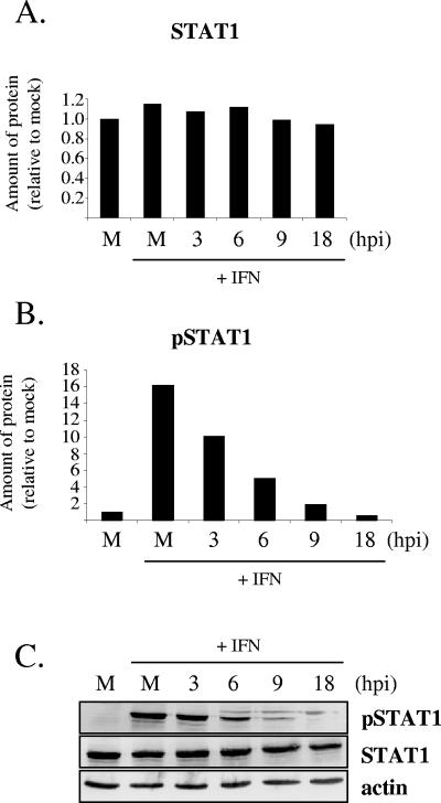
FIG. 3.
Effect of PRV Be infection on tyrosine phosphorylation of STAT1 in REF cells. Cells were either mock infected (M) or infected with PRV Be at a high MOI for various time periods before being stimulated with IFN-β for 30 min or left unstimulated. Whole-cell lysates were examined by Western blot analysis with antibodies recognizing phosphorylated (Tyr701) STAT1 (pSTAT1), STAT1, and actin. Band intensities were quantified with a phoshorimager and normalized to actin levels for each sample. The amounts of STAT1 (A) and pSTAT1 (B) in the mock-infected, unstimulated, sample were set to 100%. All other measurements are presented relative to that. Each measurement is an average of two separate experiments. A representative Western blot is shown in panel C.

