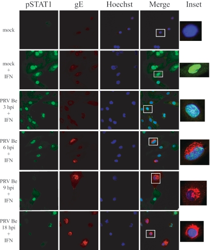
FIG. 4.
Immunofluorescent detection of STAT1 phosphorylation and nuclear translocation in PRV Be-infected REF cells. Cells were either mock-infected or infected with PRV Be at a low MOI for various time periods before being stimulated with IFN-β for 30 min or left unstimulated. Representative immunofluorescent images stained with anti-pSTAT1 and anti-gE antibodies and Hoechst DNA stain are shown. Cells outlined in the merged panels are magnified in the inset column. Image acquisition settings were kept constant for all panels.

