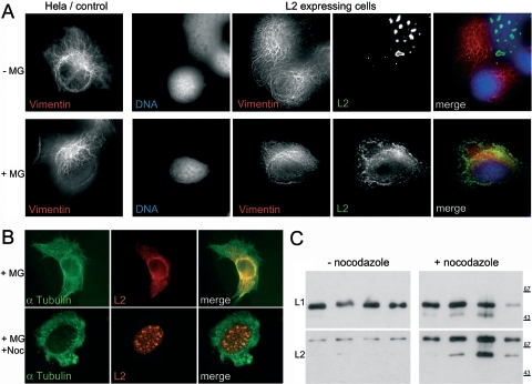
FIG. 2.
Analysis of L2 function in the presence of MG132. HPV16 L2-expressing HeLa cells were stained for vimentin (mouse monoclonal antibody; Sigma) and L2 (K18) (31) and observed by fluorescence microscopy using 3D deconvolution (A). HeLa cells were treated as described in the Fig. 1 legend in the presence or absence of nocodazole and stained with L2- and α-tubulin-specific (Sigma) antibodies (B). VLPs obtained from HeLa cells treated with MG132 in the absence or presence of nocodazole were subjected to sucrose gradient centrifugation. Peak fractions were analyzed for L2 incorporation by Western blotting using L1- and L2-specific antibodies L1-7 and L2-1, respectively (23, 32) (C).

