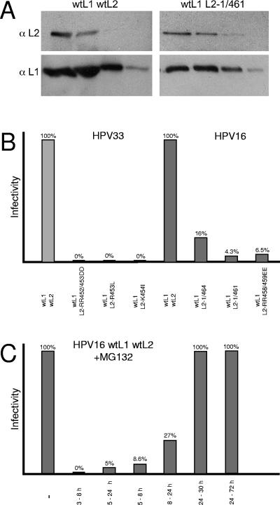
FIG. 4.
Infectivity of mutant HPV33 and HPV16 pseudovirions. Peak fractions of Optiprep gradients loaded with wild-type (wt) and mutant pseudovirions, respectively, were analyzed for the presence of L1 and L2 by Western blotting (A). Cells were infected with equal amounts of wild-type or mutant pseudovirions. Infectious events were monitored 72 h postinfection by counting cells exhibiting green nuclear fluorescence (B). Cells were infected with wild-type HPV16 pseudovirions in the absence or presence of 10 μM MG132 at the time periods indicated (C).

