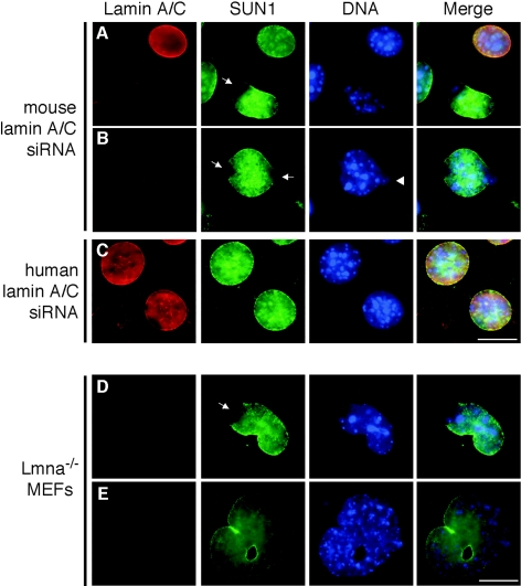
FIG. 5.
Lamin A/C is not required for localization of SUN1 to the NE. Subcellular localization of SUN1 in RNA interference-treated NIH 3T3 (A to C) or Lmna knockout MEF cells (D and E). NIH 3T3 cells were transfected with a mouse (A and B) or a control human (C) lamin A/C siRNA and processed for immunofluorescence microscopy 48 h after transfection. Cells were stained with anti-lamin A/C XB10 (red) and anti-SUN1 (green) antibodies. Hoechst staining of DNA is in blue. Arrows indicate disrupted SUN1 localization at the poles of nuclei. The arrowhead indicates apparent leakage of DNA from the nucleus. Scale bars, 10 μm.

