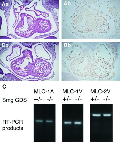
Figure 4.
Normal proliferation and differentiation of cardiomyocytes in the Smg GDS−/− heart. Transverse sections through the heart of embryos at 11.5 dpc are shown. (Aa and Ba) HE staining of the Smg GDS+/− heart (Aa) and the Smg GDS−/− heart (Ba). (Ab and Bb) BrdU incorporation into proliferating cardiomyocytes was measured by immunohistochemical analysis. Sections of Smg GDS+/− (Ab) and Smg GDS−/− (Bb) embryos at 13.5 dpc were stained with a BrdU-specific antibody, and BrdU-positive cells per field were scored. (C) RT-PCR products derived from Smg GDS+/− and Smg GDS−/− heart mRNAs at 11.5 dpc were electrophoresed and stained with ethidium bromide. Primers were specific for the MLC-1A, -1V, and -2V genes.

