Figure 5.
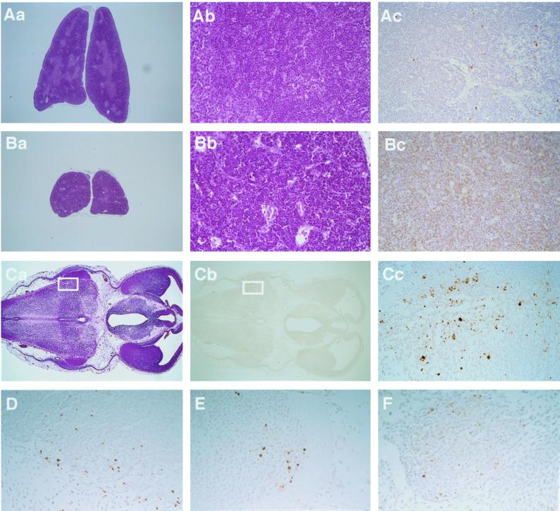
Enhanced apoptosis in the Smg GDS−/− thymus at 1 d after birth and increased neuronal cell death in the Smg GDS−/− embryo. (Aa and Ba) Sections of the wild-type thymus (Aa) and the Smg GDS−/− thymus (Ba) were prepared from littermate mice and stained with HE (20×). The Smg GDS−/− thymus was one-third in size of the wild-type thymus. (Ab and Bb) HE staining of the wild-type thymus (Ab) and the Smg GDS−/− thymus (Bb) at high-power (200×) magnification. Numerous pyknotic nuclei were found in Smg GDS−/− thmocytes compared with the wild-type thymocytes. (Ac and Bc) TUNEL staining of the wild-type thymus (Ac) and the Smg GDS−/− thymus (Bc). TUNEL-positive thymocytes were increased in the Smg GDS−/− thymus. (Ca and Cb) Transverse sections through the third ventricle and the diencephalon of the Smg GDS−/− embryo at 12.5 dpc. (Ca) HE staining of the Smg GDS−/− brain (20×). (Cb) TUNEL staining of the Smg GDS−/− brain (20×). (Cc–F) High-power (200×) magnification. TUNEL-positive neuronal cells were increased in the medulla oblongata (Cc; boxed in Ca and Cb), the trigeminal ganglia (D), the spinal cord (E), and the dorsal root ganglia (F).

