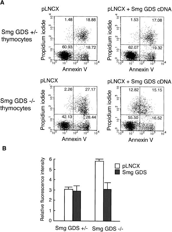
Figure 7.
Rescue of etoposide-induced apoptosis by the Smg GDS cDNA exogenously expressed in Smg GDS−/− thymocytes. (A) Cultures of 1 × 10⁶ thymocytes derived from newborn Smg GDS+/− (upper panels) and Smg GDS−/− (lower panels) mice were transduced with empty retroviral vector pLNCX (left panels) and transduced with pLNCX expressing Smg GDS cDNA (right panels). Retrovirus-treated thymocytes were then stimulated for 4 h with etoposide to induce apoptosis, stained with propidium iodide and annexin V-FITC, and analyzed by flow cytometry. The percentage of the total gated cells analyzed is given within the panels. (B) Caspase-3 activity was determined as in Figure 6. Mean values are shown for five independent experiments using Smg GDS+/− and Smg GDS−/− thymocytes after treatment with the empty retroviral vector pLNCX (open boxes) and pLNCX expressing Smg GDS cDNA (shaded boxes) and treatment with etoposide for 4 h.

