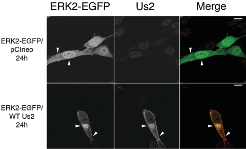
FIG. 3.
Us2 expression alters the subcellular localization of ERK2-EGFP. NIH 3T3 cells were cotransfected with the plasmid combinations indicated on the left of the figure. Twenty-four hours after transfection, cells were stained for Us2. On merged images, the ERK2-EGFP signal is green, and the Us2 signal is red. pCIneo is an empty expression plasmid and has no effect on ERK2-EGFP localization. Arrowheads indicate points of interest. Images were obtained using a Zeiss 510 confocal laser scanning microscope. Scale bars are 15 μm.

