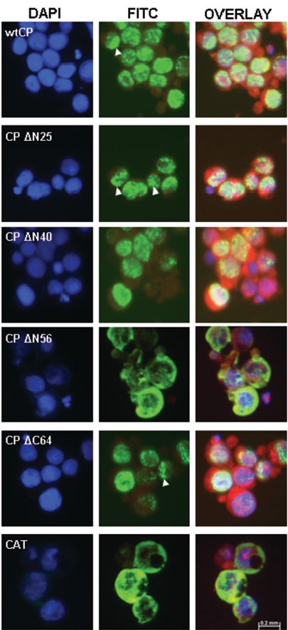
FIG. 2.
Subcellular distribution of BFDV capsid protein. Insect cells were infected with recombinant baculovirus and probed with a monoclonal antibody directed against the N-terminal His6 affinity tag. Antibody binding was followed by a fluorescein isothiocyanate (FITC)-conjugated secondary antibody (green). DAPI stain was used to define nuclei (blue), while membranes were visualized with Evans blue (red). Merged images (overlay) are also shown. Arrowheads indicate specific examples of the punctate perinuclear distribution of the relevant recombinant proteins.

