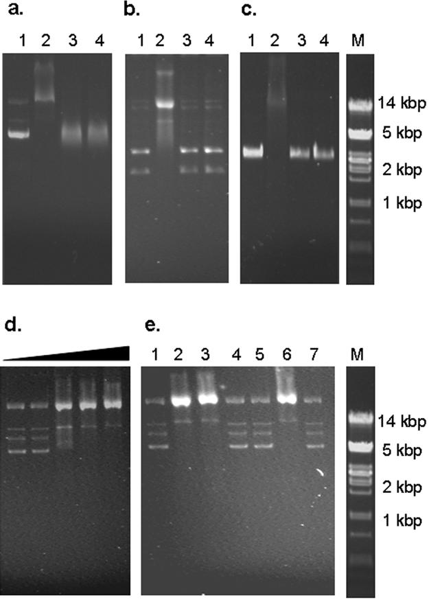
FIG. 3.

Electrophoretic mobility shift analysis of the interaction of recombinant BFDV CP with various DNA samples. (a) Plasmid preparations containing the BFDV genome, (b) plasmid preparations of pUC18, and (c) phage M13 ssDNA DNA samples were incubated in the absence of protein (lane 1), in the presence of purified wt CP (lane 2), and in the presence of purified CAT (lane 3). To confirm that the observed retardation of the DNA fragment was indeed caused by bound proteins, 1 μg/ml proteinase K was added to the samples containing purified CP (lane 4). (d) Increasing amounts of His-tagged CP were incubated with BFDV DNA. Lane 1, DNA only; lane 2, 10 ng; lane 3, 20 ng; lane 4, 50 ng; lane 5, 500 ng. (e) The interaction of each of the truncated variants of the BFDV CP with BFDV DNA was assessed in a similar manner. Lane 1, DNA only, lane 2, wt CP; lane 3, CP ΔN25; lane 4, CP ΔN40; lane 5, CP ΔN56; lane 6, CP ΔC64; lane 7, CAT. Lanes M, molecular size markers.
