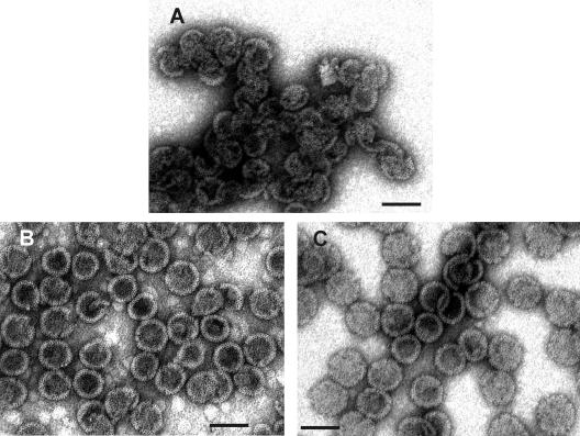
FIG. 3.
Transmission EM images of negatively stained M-PMV particles assembled from ΔProCANC protein. (A) Particles assembled from ΔProCANC protein without addition of nucleic acids. In this case, about 80% represented fragments not fully closed and interconnected particles. (B) Particles assembled from ΔProCANC protein and bacteriophage MS2 RNA at a ratio of 10:1 (wt/wt). (C) Particles assembled from ΔProCANC protein and 16S plus 32S rRNA of E. coli in a ratio of 10:1 (wt/wt). More than 90% of particles assembled in the presence of both MS2 RNA and rRNA were fully closed spheres. The average diameter of all assembled spheres was 75 nm ± 4 nm. Approximately 15%, 65%, and 75% of protein was efficiently assembled into particles in the absence of nucleic acid and in the presence of MS2 RNA and rRNA, respectively. The gross estimate (based on the number of molecules in the particle and the protein amount) is that 100% of assembled protein corresponds to 3 × 10¹¹ particles. All assembly reactions were performed as mentioned in Materials and Methods. Bar, 100 nm.

