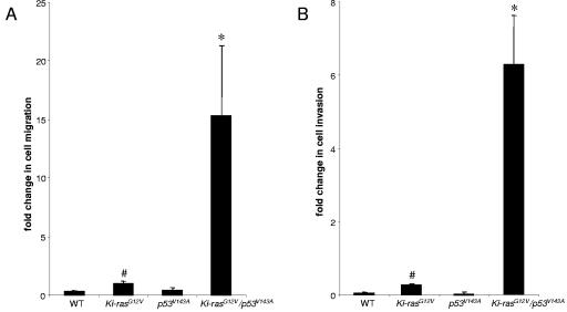
FIG. 1.
Ki-RASG12V/p53V143A-PDC display increased migration and invasion. (A) Pancreatic ductal cells representing the different genotypes were seeded on noncoated Boyden chamber membranes for migration assays. After 24 h, the cells that did not migrate were removed using cotton swabs. The migrating cells were stained and quantified by averaging 10 individual fields. (B) Pancreatic ductal cells representing the different genotypes were seeded on Matrigel-coated Boyden chamber membranes for invasion assays. After 24 h, the cells that did not invade were removed while the invading cells were stained and quantified by averaging 10 individual fields. Symbols: *, P < 0.02 (Ki-RASG12V/p53V143A-PDC versus Ki-RASG12V-PDC or p53V143A-PDC), #, P < 0.05 (Ki-RASG12V-PDC versus WT-PDC). The data were obtained from three independent experiments performed in duplicate.

