FIG. 4.
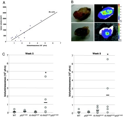
In vivo bioluminescence imaging of WT-PDC, p53V143A-PDC, Ki-RASG12V-PDC, and Ki-RASG12V/p53V143A-PDC stably transduced with a pFB-neo/luc retroviral vector. Irradiated immunodeficient mice received subcutaneous injections of 1 × 10⁷ cells suspended in a 1:1 ratio of Matrigel/DMEM/F12 full medium. Animals were imaged weekly for 10 weeks to monitor tumor growth. (A) Correlation between tumor volume (mm3) and bioluminescence signal (photons per second [ph/s]) at various time points of tumor development in mice injected with WT-luc-PDC, p53V143A-luc-PDC, Ki-RASG12V-luc-PDC, and Ki-RASG12V/p53V143A-luc-PDC. Data are shown for individual mice at 1 to 8 weeks after subcutaneous injections (R2 = 0.72). (B) After 10 weeks, ex vivo imaging of excised tumor tissues confirmed the presence of luciferase activity arising only from the Ki-RASG12V/p53V143A-luc-PDC. Bioluminescence was measured 20 min after injection of the d-luciferin substrate. (C) Bioluminescence and growth of subcutaneous luc-PDC tumors are shown for week 5 and week 8 after subcutaneous injections of WT-luc-PDC (n = 6), p53V143A-luc-PDC (n = 6), Ki-RASG12V-luc-PDC (n = 7), and Ki-RASG12V/p53V143A-luc-PDC (n = 5), with a horizontal bar representing the mean value for each cell line. n, number of tumors considered for this experiment. *, P was <0.05 for Ki-RASG12V/p53V143A-PDC versus WT-luc-PDC, p53V143A-luc-PDC, or Ki-RASG12V-luc-PDC.

