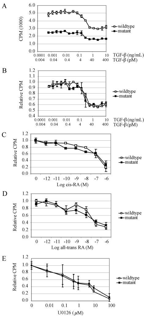
FIG. 6.
Analysis of MEF proliferation in response to TGF-β, 9-cis-RA, all-trans-RA, and U0126. (A) Primary Tgif+/+ and Tgif−/− MEF cells were grown in the presence of 0.6 pg/ml to 10 ng/ml TGF-β for 24 h and DNA synthesis was measured by [3H]thymidine incorporation as described in the text. Mutant MEFs displayed lower proliferation at all concentrations of TGF-β relative to wild-type MEFs. (B) Results shown in panel A were normalized to the maximum value of each genotype, since mutant MEFs consistently showed reduced proliferation. The proliferation inhibitory responses of wild-type and mutant MEFs were similar at all concentrations of TGF-β. (C to D) Fresh 9-cis-RA (C) and all-trans-RA (D) were added to MEFs on a daily basis for 5 days, and [3H]thymidine incorporation was performed on day 6. Results were normalized as described for panel B. The proliferative inhibitory responses to 9-cis-RA and all-trans-RA were similar for wild-type and mutant MEFs. (E) Concentrations ranging from 0.01 to 100 μM U0126 were added to MEFs and [3H]thymidine incorporation was performed after 24 h of culture. The proliferative inhibitory responses to U0126 were similar for wild-type and mutant MEFs. All results are from representative experiments. For each, three experiments were performed in triplicate using cells derived from independent Tgif+/+ and Tgif−/− embryos. Error bars represent standard deviations of the means. CPM, counts per minute.

