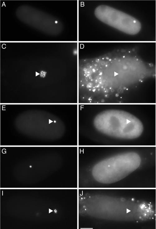
FIG. 2.
ECFP-HP1α and ECFP-SUV39H1 localize to the condensed transgenic locus. Cells were transiently transfected with combinations of plasmids encoding proteins as follows: EYFP-LacR and ECFP-HP1α (A and B) (silent); EYFP-LacR-VP16, ECFP-HP1α, and hTet-Off (C and D) (double activation); EYFP-LacR and ECFP-HP1α V22M (E and F) (silent); EYFP-LacR and ECFP-SUV39H1 (G and H) (silent); and EYFP-LacR, ECFP-SUV39H1, and hTet-Off (I and J) (single activation). The left panels show the transgenic loci, and the right panels show the distribution patterns of the ECFP fusion proteins. The cytoplasmic punctate fluorescence (D and J) represents peroxisomal CFP signals in the activated cells. The triangles indicate the sites of the transgenic loci. Scale bar, 5 μm.

