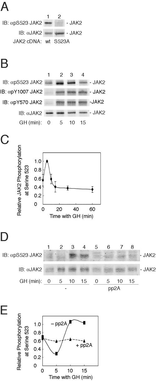
FIG. 2.
GH promotes the phosphorylation of JAK2 at serine 523. (A) 293T cells transfected with cDNA (1 μg) encoding either JAK2 (lane 1) or JAK2 S523A (lane 2) were lysed. The lysates were immunoblotted (IB) with αpSer523 JAK2 (upper panel) or αJAK2 (lower panel) (n = 4). wt, wild type. (B) 3T3-F442A cells were treated with vehicle or with 500 ng GH/ml for 5, 10, or 15 min. The cells were lysed, and lysates were immunoblotted (IB) with αpS523 JAK2 (top panel), αpY1007/1008 JAK2 (second panel), αpY570 JAK2 (third panel), or αJAK2 (bottom panel). (C) Replicates for the experiment shown in panel B were normalized to levels of JAK2. Means ± standard errors of the means are shown for n = 6. (D) 3T3-F442A cells were treated with vehicle (lanes 1 and 5) or with 500 ng GH/ml (lanes 2 to 4 and 6 to 8). Cells were lysed, and lysates were incubated without (lanes 1 to 4) or with (lanes 5 to 8) 0.7 U pp2A at 37°C for 60 min. Cell lysates were immunoblotted with either αpS523 JAK2 (top panel) or αJAK2 (bottom panel). The migration of JAK2 is indicated. (E) Results from the experiment shown in panel D were quantified and normalized to levels of JAK2.

