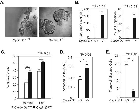
FIG. 4.
Cyclin D1 regulates cellular adhesion and migration. (A) cyclin D1−/− MEFs display increased apposition detected by interference reflection microscopy (60×). Scale bar, 20 μm. (B) Quantitation of dark area pixel and % cell apposition (the ratio of dark area pixel to the total area pixel) of cyclin D1+/+ and cyclin D1−/− MEFs by ImageJ software (http://rsb.info.nih.gov/ij/). (C) cyclin D1−/− MEFs spread more rapidly than cyclin D1+/+ MEFs. cyclin D1+/+ and cyclin D1−/− MEFs were assessed by enumerating dark and bright cells under phase contrast. Dark cells were considered to be spread, and bright cells were considered to be unspread. The mean ± standard error of the number of spread cells is shown at the time points 30 min and 1 h after plating. (D) cyclin D1−/− MEFs are more adherent than wild-type MEFs. A total of 20,000 wild-type and cyclin D1−/− MEFs were seeded in a 96-well plate coated with 10 μg/ml fibronectin. After 30 min, plates were washed two times with PBS, fixed, and stained with crystal violet, and the absorbance was read at 550 nm (A550). (E) cyclin D1−/− MEFs migrate more slowly than wild-type MEFs. A total of 40,000 cells were seeded in a fibronectin-coated Transwell. Cellular migration was determined by the cell number at the bottom of the Transwell plate as described in Materials and Methods.

