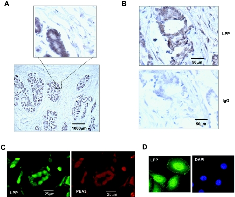
FIG. 2.
LPP expression in normal and cancerous breast tissue. (A) Immunohistochemical analysis of LPP protein expression in normal human breast tissue sections. An enlarged part of the image is shown to illustrate expression in the ductal epithelial cells. (B) LPP expression from human tissue derived from patients with invasive ductal carcinomas. LPP expression is revealed by brown staining. A control IgG-stained adjacent section is shown in the bottom panel. (C) Immunofluorescence analysis of LPP expression (green, fluorescein isothiocyanate) and PEA3 expression (red, tetramethyl rhodamine isothiocyanate) in ductal carcinoma samples. (D) Confocal analysis of LPP expression (fluorescein isothiocyanate; green) in MDA-MB-231 cells. Nuclei are visualized with DAPI staining.

