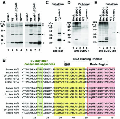
FIG. 1.
SUMO-2/3 is conjugated to MafG in the bone marrow. (A) Immunoblot analysis of whole-cell lysates from bone marrows reacted with anti-small Maf antibody. The bone marrow samples were prepared from mafG-null mutant mice (lanes 1 and 5), wild-type mice (lanes 2 and 6), G1HRD-MafG transgenic mice in a mafG-null mutant background (lane 3) or wild-type background (lane 4), and G1HRD-MafK transgenic mice in a mafG-null background (lane 7) or wild-type background (lane 8). (B) Alignments of the N-terminal halves of small Maf proteins found in human, mouse, chicken, and zebra fish. Amino acids comprising the extended homology region (EHR) and basic region are boxed. The conserved sumoylation consensus site is also boxed. (C to E) Nickel pull-down samples of the bone marrows from wild-type mouse and G1HRD-His-MafG mouse (line 11) reacted with anti-MafG antibody (C, lanes 1 and 2), anti-SUMO-1 antibody (D, lanes 4 and 5), or anti-SUMO-2/3 antibody (E, lanes 4 and 5). The 239T cell lysate and those containing overexpressed hemagglutinin (HA)-tagged SUMO-1 and SUMO-2 were used to confirm antibody specificity (lanes 1 to 3 in panels D and E). White and black arrowheads indicate small Maf with or without modification, respectively (A, C, and E). Asterisks indicate nonspecific signals (A).

