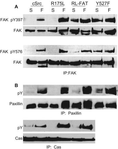
FIG. 5.
Adhesion signaling of SYF cells reconstituted with cSrc, SrcR175L, SrcY527F, and SrcR175L-FAT. The figure shows Western blots indicating phosphorylation levels of FAK at tyrosine residues 397 and 576 (A), as well as total phosphotyrosine of paxillin and Cas (B) in suspension (S) and after 1 h of spreading on fibronectin (F). (A) Phosphorylation of FAK at residue Y397 in whole-cell lysates and Y576 in FAK immunoprecipitates of SYF cells and cells reexpressing cSrc and Src mutants. Membranes were reprobed with anti-FAK MAb for normalization. (B) Reaction of antiphosphotyrosine antibody (RC20) to immunoprecipitates of Cas and paxillin from SFK-null fibroblasts reconstituted with wild-type Src and Src mutants.

