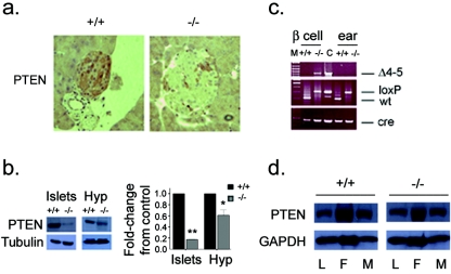
FIG. 1.
Tissue-specific deletion of Pten. (a) Immunohistochemistry showing PTEN deletion in β cells (original magnification, ×25). (b) Western blots (left panels) and quantification of Western blot signal (right panel) showing decreased expression of PTEN in isolated islets and the hypothalamus (Hyp). Residual signal in islets is likely due to the presence of non-β cells. *, P = 0.011; **, P < 0.005. The error bar indicates the standard error of the mean. (c) PCR analysis of Cre-mediated recombination of the Pten locus (Δ4-5, top) in β cells and ear tissue and genotyping for Pten-loxP allele (middle) and Cre (bottom). M, marker; C, control mammary gland tumor; wt, wild type. (d) Expression of PTEN in liver (L), fat (F), and muscle (M) is unchanged. +/+, RIPcre+ Pten+/+ mice; −/−, RIPcre+ Ptenfl/fl mice.

