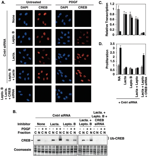
FIG. 3.
Leptomycin B and lactacystin inhibit PDGF-induced CREB depletion, loss of CREB transcriptional activity, and SMC proliferation. Rat PA SMCs were transfected with nonspecific (Cntrl) or CREB-specific double-stranded siRNAs as indicated and incubated overnight in DMEM containing 15% FCS. The cells were transferred to DMEM containing 0.2% FCS for 24 h. The cells were then treated with 100 nM leptomycin B (Lepto. B) and/or 500 nM lactacystin (Lacta.) for 30 min prior to the addition of 25 ng/ml PDGF. Fresh medium containing PDGF and inhibitors was added every 24 h for 72 h. (A) Cells were fixed, subjected to immunostaining with a CREB-specific antibody, and counterstained with DAPI to indicate nuclei. Magnification, ×400. (B) Cytosolic (“C”) and nuclear (“N”) extracts were prepared and separated on 10% polyacrylamide-SDS gels and transferred to PVDF membranes. Western blotting was performed with a CREB-specific antibody. The positions of CREB and ubiquitinated CREB (Ub-CREB) are indicated. A representative Coomassie blue-stained blot is shown as a loading control. (C) Prior to being treated with PDGF and inhibitors, cells were transfected with a plasmid containing a CREB-responsive promoter linked to the firefly luciferase gene (pCRESV-Luc). Cells were cotransfected with the internal control vector pRL-SV40 to correct for transfection efficiency. Following treatment with PDGF and inhibitors, firefly luciferase levels were determined as an index of CREB transcriptional activity. The figure shows transcription relative to levels measured in cells not treated with PDGF and inhibitors. Untreated, cross-hatched bars; PDGF treated, solid bars. (D) SMC proliferation was measured with CellTiter 96 AQ reagents as described in Materials and Methods. Proliferation is expressed relative to absorbances measured with cells not treated with PDGF or inhibitors. Untreated, cross-hatched bars; PDGF treated, solid bars.

