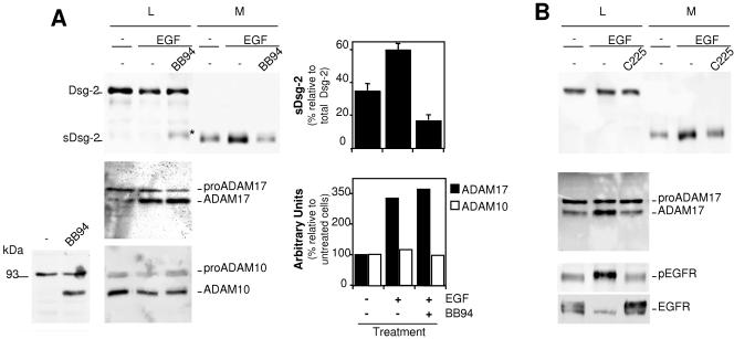
FIG. 5.
Analysis of the shedding of Dsg-2 in cells treated with EGF. A. Parental A431 cells were treated with or without EGF and BB-94 as indicated and lysed. Cell lysates (L) and medium samples (M) were analyzed by Western blotting with antibodies against Dsg-2, ADAM17, or ADAM10 as indicated. Note that when a lysis buffer without BB-94 is used, only pro-ADAM10 is detected in cell lysates from A431 cells (bottom left). Addition of BB-94 allows the detection of mature ADAM10 (bottom left), and thus a buffer containing this metalloprotease inhibitor was used in the right bottom panel. Western blots were quantified, and the averages from three independent experiments ± standard deviations or the averages from two experiments are shown. B. A431 cells were treated with or without EGF and C225 (a monoclonal antibody that blocks the activation of the EGFR) for 48 h. Cell lysates and medium samples were analyzed by Western blotting with antibodies against Dsg-2, ADAM17, phospho-EGFR, or EGFR as indicated.

