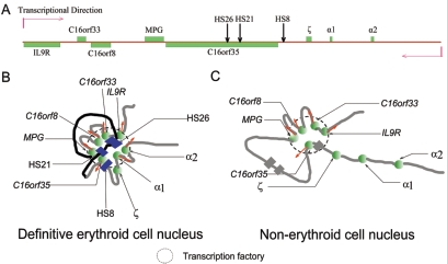
FIG. 6.
Schematic representation of the linear structure in genome (A) and the putative three-dimensional structure of a large chromatin region in definitive erythroid cells (B) and nonerythroid cells (C). In definitive erythroid cells, active α-globin genes are in close proximity with the upstream HSs to form an α-ACH, similar to β-ACH. Moreover, mouse α-ACH colocalizes with the upstream housekeeping genes, which may form a subcompartment in the nucleus like a “transcription factory” where the concentrated RNA polymerase may be shared by a particular group of genes (B). In nonerythroid cells (C), the repressed mouse α-globin genes are separated from the congregated housekeeping genes, which retain a “transcription factory” shared by these housekeeping genes. The symbols in panel A are as described in the legend to Fig. 1. In panels B and C, the green spheres represent protein complexes on gene promoters. The gray and black strings represent chromatin. The red arrows indicate transcription direction. In panel B, the blue boxes represent protein complexes on distant erythroid-specific HSs. In panel C, the gray boxes show the positions of erythroid cell-specific HSs.

