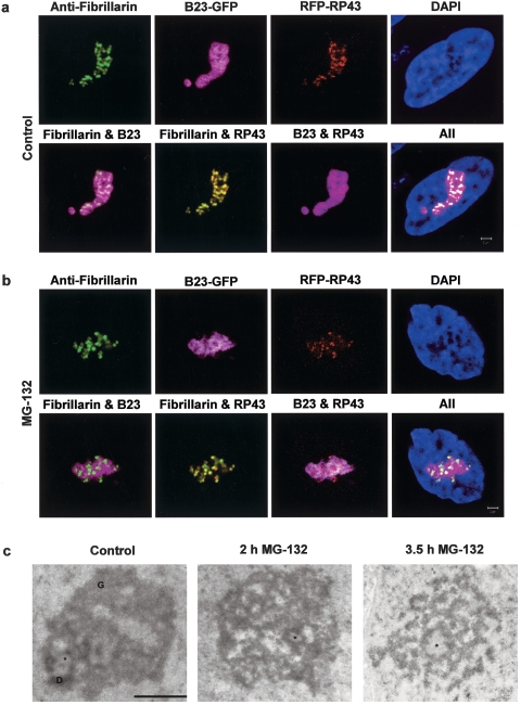
FIG. 2.
Proteasome inhibition by MG-132 and lactacystin induces morphological changes in the nucleolus. Treatment of B23-GFP transfected HeLa cells with 100 μM MG-132 or 50 μM lactacystin (data not shown) for 2 h changes the relative distributions of B23 and fibrillarin, as well as B23 and RP43 (compare images in the second rows of panels a and b). Scale bar, 2 μm. The normal ultrastructural organization of the nucleolus is affected after treatment with 100 μM MG-132 (c). Although structural counterparts of fibrillar centers (FC, asterisk) are observed both after 2 h and after 3.5 h of treatment, granular components (G) and dense fibrillar components (D) gradually disintegrate. Scale bar, 1 μm.

