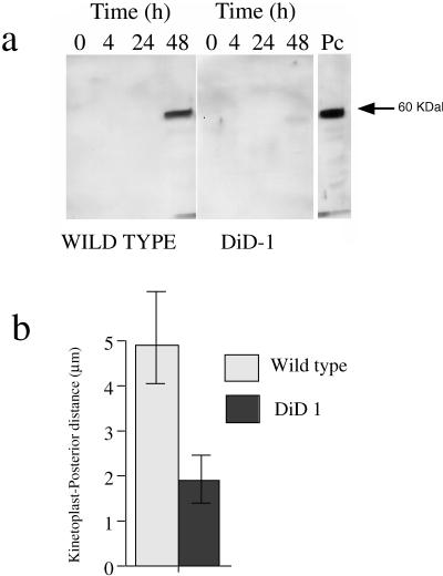
Figure 3.
Glycosomal development and morphological restructuring of DiD-1. (a) Glycosomal development of DiD-1 cells 4, 24, and 48 h after culture at 27°C in 6 mM cis-aconitate. The left panel shows equal protein loadings of the wild-type population undergoing differentiation, and the right panel shows DiD-1 samples under the same conditions. Each blot was probed with a rat anti-PEPCK antibody (a kind gift from Prof. T. Seebeck, University of Bern, Bern, Switzerland). A procyclic protein sample is shown at the extreme right. (b). Quantitative analysis of the relative distance between the kinetoplast and cell posterior in wild-type or DiD-1 populations 24 h after incubation in differentiation conditions. n = 100 cells for each cell type.

